How can AFM be used for biological research?
Atomic force microscopy for biofilms, spores, and more.
We recently had the privilege of hosting Professor Joel Weadge and PhD student Emily Rodriguez from Wilfrid Laurier University at ICSPI’s facilities for a morning visit and some sample scanning! They brought in dried Gram-positive microbiological samples on glass slides and filter paper related to their research on biofilms and the latter’s role in bacterial survival and propagation.
Normally, getting scans like this can take over a day and may involve sending samples to a core facility. But, using the Redux Atomic Force Microscope, we achieved these nanoscale results across multiple samples on a lab benchtop in a single morning.
Join us on this blog post as we discuss what we were able to see together using AFM and the new insights into biological research we gained!
What can we see under optical microscope?
Using our Redux AFM, we were first able to take a look at the samples under the instrument’s integrated optical microscope and navigate to various areas of interest where bacterial density was both higher and lower using the motorized XY sample stage.
Using AFM to identify control vs. mutant bacteria colonies
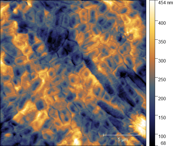
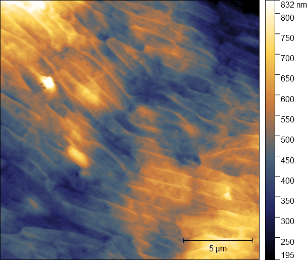
Some samples represented wild-type control group bacteria whereas others were genetically mutated colonies. These mutant strains were modified to be less efficient at forming biofilms. We were able to hydrate both types of these samples and scan them using our AFM.
In these scans of highly concentrated bacterial areas, AFM let us easily discern the differences between these two cell groups. Through these topographical images, one can see how the wild strain (left) remains in its tubular cell form, while the mutant strain (right) faces more environmental stress due to impaired biofilm formation and as a result has more lysed cells and forms spores more readily for survival.
Imaging bacterial spore structures
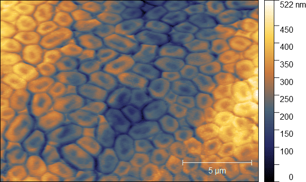
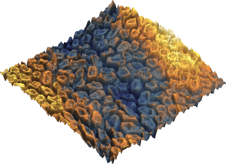
Navigating to areas of lower bacterial density on the samples, where spore formation is a necessity for survival and self-assembly easily visible. we able to gather more data and directly image the bacterial spores and their formed structures.
Using these 2D (left) and 3D (right) maps, we were even able to measure the spore coating thickness (~50 nm) and observe the spore core itself.
What can researchers do with this?
By studying morphological changes through performing nanoscale cell imaging, we are able to confirm structural changes resulting from genetic modification and environmental influences.
AFM in particular has advantages over other common complementary techniques such as cryo-electron microscopy by being significantly easier and faster to use as well as changing the game by allowing hydrated sample scanning in ambient conditions.
With this, we can rapidly establish clear structure-property relationships and drive new research processes.
When we better understand the behaviour of bacteria in biofilms and spores, we can gain greater insights into sanitation and preventing the spread of harmful bacteria in a variety of applications ranging from medical device design to surgical outcomes.
Want to learn even more about AFMs for biological research?
If you’re interested in learning more about how AFM can transform your biological research, check out the Publications page on our website to see what others have achieved using our AFM systems.
We would also love to directly connect and learn more about your work! Feel free to reach out to us at our Contact Us page.
Interested in learning how AFM can help you?
Speak with an application engineer today to see how AFM can help with your research and process development.


