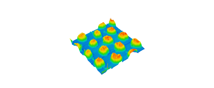
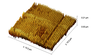
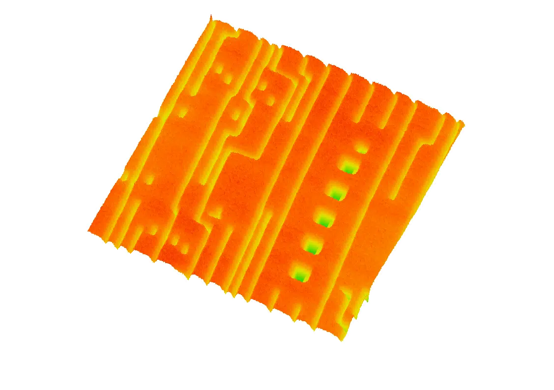
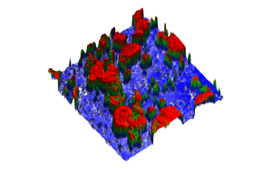
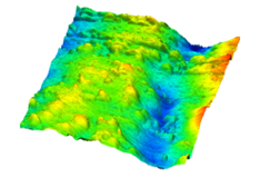
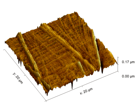
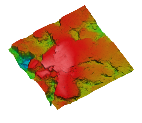
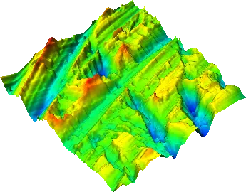
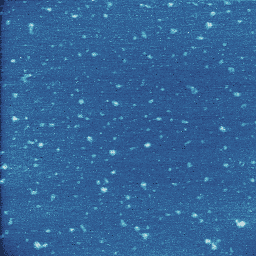
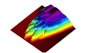
Atomic force microscope images Atomic force microscope images Browse our gallery of atomic force microscope images. All the images in this gallery were collected using ICSPI’s AFM instruments.For a deep-dive into any particular application, check out our Application Notes page.For publications that have used ICSPI’s AFM instruments, check out our Publications page. Semiconductors and Microfabrication Photonic grating made from silicon. Pillars are on a 3 micron pitch. Topography scan. Structures made on polymer using nanoimprint lithography. 100 nm tall structures on a 100 nm pitch. Topography scan in 3D view. Photoresist on silicon wafer. Topography scan in 3D view Polymers and Composites Phase image of silica-polymer composite. Red areas show the hard silica particles and blue areas are the soft polymer matrix. 10 micron scan. Acrylo-butadiene-styrene (ABS) terpolymer. Topography image. Linear low-density polyethylene (LLDPE) polymer sheet. The roughness of the sheet was correlated to poor adhesive performance. Metals, Minerals and Ceramics Aluminum nitride. Topography scan. 3d-printed titanium-aluminum alloy polished to mirror finish. Ra = 13.6 nm Polished steel with 9-micron sized diamond polish. Image courtesy of Akasel A/S. Biology, Biotechnology and Life Science Human skin cells with keratin filaments. Topography scan. Extracellular vesicles. Vesicles are 20 - 30 nm tall. Topography scan. Rose petal. Topography scan. Have an application in mind? Speak with an Application Engineer today to see if AFM can help. Contact Us