AFM for Biology and Life Sciences
Common applications of AFM in biology and life science
Atomic Force Microscopy (AFM) is a versatile imaging technique for biology and life science. AFM can non-destructively collect high-resolution 3D surface images of biological material at the nano- and microscales. Applications in biology and life science include imaging of bacteria, viruses, fungi, proteins, tissues, sera, as well as functionalized surfaces for biotechnology applications.
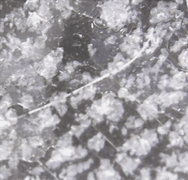
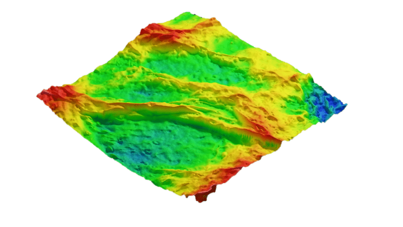
In applications such as examining skin samples, AFM offers significantly higher magnification and resolution than optical microscopy. Structures like keratin fibers and corneocytes become clearly visible, thanks to AFM’s capability to produce three-dimensional data. This high-resolution three-dimensional imaging is superior to confocal microscopy, which has limited Z-axis resolution, thus providing only a fraction of the detail that AFM can achieve with its sub-nanometer Z-axis resolution.
Such detailed data is crucial for identifying specific nanoscale features in biological samples, potentially enabling non-invasive biopsies for diagnosing skin diseases or studying the structural coloration in butterfly wings. Additionally, AFM can analyze biopolymers like collagen, offering insights into their structural arrangement and properties.


Benefits of AFM for biology and life science
AFM can scan virtually any solid sample with minimal sample preparation and without destroying the sample. This is a stark contrast to Scanning Electron Microscopy (SEM) and Transmission Electron Microscopy (TEM), which require extensive sample preparation such as metal coating, and/or cross-sectioning, potentially altering or damaging the sample.
Want to learn more?
Read our one-page application note that covers multiple samples of biological origin: keratin filaments on skin cells, butterfly wings, and collagen.
Speak with an expert
Interested to learn more about how AFM can help you with your biological samples?
